Monday, 23 December 2024

Focused Shockwave Therapy

Focused Shockwave Therapy (FSWT) is a medical treatment that uses high-energy acoustic waves to promote healing, reduce pain, and improve function in various musculoskeletal conditions. It is commonly used in orthopedics, sports medicine, and physical therapy.

How it Works:

  1. Energy Delivery: High-energy waves are focused precisely on a specific area of the body.
  2. Mechanical Effects: These waves create microtrauma or stress in the tissues, stimulating the body's natural healing processes.
  3. Biological Response:
    • Increases blood flow to the treated area.
    • Stimulates the production of growth factors.
    • Promotes the regeneration of tissues like tendons, ligaments, and bones.
    • Reduces calcifications and scar tissue.

Conditions Treated:

  • Plantar fasciitis
  • Achilles tendinopathy
  • Patellar tendinopathy
  • Lateral epicondylitis (tennis elbow)
  • Medial epicondylitis (golfer's elbow)
  • Calcific shoulder tendinopathy
  • Stress fractures and delayed bone healing
  • Chronic myofascial pain
  • Ankle instability

Benefits:

  • Non-invasive
  • No need for anesthesia
  • Minimal side effects
  • Short treatment sessions (10-30 minutes)
  • Can be an alternative to surgery in some cases

Procedure:

  1. The patient is positioned comfortably.
  2. A gel is applied to the skin to improve acoustic wave transmission.
  3. A handheld device delivers focused waves to the targeted area.

Side Effects:

  • Temporary pain or discomfort
  • Redness or swelling in the treated area
  • Bruising (rare)

Focused vs. Radial Shockwave Therapy:

  • Focused Shockwave Therapy: High-energy, targeted, and penetrates deeper into tissues. Suitable for conditions like bone healing or deeper tendinopathies.
  • Radial Shockwave Therapy: Lower energy, disperses over a broader area, often used for surface-level conditions.

If you're considering FSWT, consult with a healthcare professional to determine if it's appropriate for your specific condition.

2022 Study on the Effectiveness of Focused Shockwave Therapy for ankle instability.

http://www.kptjournal.org/journal/view.html?doi=10.18857/jkpt.2022.34.3.91

Friday, 28 January 2022

Georgetown Ankle Ligament Reconstruction Protocol +/- Arthrex Internal Brace 2025

Dr. Christopher Lu MBchB FRCSC / Darryl Yardley M.Sc.PT
Ankle ligament reconstruction post-op protocol


PRE-OPERATIVE PHYSIOTHERAPY:


- Gait training, advice on edema control and pain management: rest, elevation, cold therapy
- Ankle strengthening; Proprioception
- Assess with AOFAS Ankle/Hindfoot Scale
- Learn how to use - crutches, knee scooter, cast protector, cast boot and cold therapy unit
- Please Call Restore Physiotherapy Georgetown - 905-702-1840 to book your pre-operative physiotherapy appointment.
- Post-operative physiotherapy can be completed either at Restore Physio Georgetown or a reputable physiotherapy clinic near your home.
- Post-operative in home physiotherapy is also available with: www.therapia.com


0 - 2 WEEKS:


- Non-weight wearing in below knee fiberglass back slab. Please note back slab is open on 3 sides this allows use of cold therapy wrap. Although you are technically non-weight bearing - not allowed to walk on the leg, you are allowed to stand and put pressure on the foot, ie. when brushing your teeth, sitting in a chair.
- Leg elevation and cold therapy for edema control
- Mobilize with crutches, knee scooter, iwalk. Please note the knee scooter is the safest option for extended mobilization.
- Keep cast boot and dry - use cast protector in shower
- Wound check at 2 weeks post-op
- Toe curls, toe spreads / extension
- Take aspirin 81 mg or 325 mg - once a day for DVT prophylaxis - if there are no contraindications or allergies.


2 - 6 WEEKS:

- Attend fracture clinic for follow-up appointment, back slab removed, wound examined.
- Immobilized in short leg fiberglass cast for a further 4 weeks. Please note this option has the lowest rate of wound breakdown / infection - approximately 1%. You have the option of transitioning to a long leg cast boot at this point in time, however this has a higher wound complication rate - approximately 20%.
- Continue to mobilize with crutches, knee scooter, iwalk.
- Limit ankle ROM to prevent wound break down.
- Keep cast clean and dry - use cast protector in shower
- Toe curls, toe spreads / extension
- Leg lifts and extension to keep quads strong.
- Take aspirin 81 mg or 325 mg - once a day for DVT prophylaxis - if there are no contraindications or allergies


6 - 12 WEEKS:


- 6 weeks - cast removed.
- Have the option of transitioning to long cast boot or ankle brace.
- Start physiotherapy at 6 weeks post-op
- ROM (Range of motion) in unilateral planes focus on dorsiflexion and progress to active exercises in protected ranges, avoid inversion, ankle traction, and tibio-fibular mobilization.
- Proprioception exercises, intrinsic muscle strengthening, start isometric eversion.
- Soft tissue treatments (plantar fascia, gastroc/soleus, achilles) and regular mobilization of intermetatarsal and midtarsal joints.
- Passive and active range of motion exercises (restore full ROM / multi-planar motions)
- Open kinetic and closed kinetic chain strengthening.
- Gradually progress closed-chain and balance / proprioception.
- Cycling, aerobic machines as tolerated.
- AVOID: ankle traction, forced plantarflexion and ankle inversion for 12 weeks


3 - 6 MONTHS:


- Progress back into athletics based upon functional status at discretion of Orthopaedic Surgeon.

- Assess with AOFAS Ankle/Hindfoot Scale  (Post-op - 3 months and 6 months)

Disclaimer:  This physiotherapy protocol is specifically designed for patients who have had their ankle instability surgery performed at Georgetown Hospital.

Diet plays an important role in post-op recovery.

- Please limit your intake of processed foods, junk food, fast food, sugar, artificial sweeteners, no soft drinks, and specifically no diet coke...
- If you are unable to dedicate time to your recovery - we do not recommend proceeding with surgery.

If you are interested in losing weight please continue reading:

- If you have time please read the book the Obesity Code by Dr. Jason Fung
https://www.amazon.ca/Obesity-Code-Unlocking-Secrets-Weight/dp/1771641258
- If you are a diabetic I would consider reading the book the Diabetes Code by Dr. Jason 
- If you are a diabetic I would recommend looking into purchasing a Free Style Libre CGM to better monitor your diabetes, the better your diabetic control the faster you will heal and this will also decrease your risk of infection.
- The Diet Doctor Website is also good to read:
https://www.dietdoctor.com/how-to-lose-weight
- There is also a good - Dr. Jason Fung support group on Facebook


Tuesday, 9 February 2016

Recommended aids to help with post-op recovery

Ortho Rx - Dr. Chris Lu Recommended Aids
1) Breg Kodiak Cold Therapy + Ankle / Foot Attachment / Game Ready / Ossur


2) Walking Aids - Crutches / Knee Scooter / iWalk 2.0


Ortho Rx - Dr. Chris Lu Recommended Aids

3) Shower Aids


Ortho Rx - Dr. Chris Lu Recommended Aids

4) Vitamin D drops - 2000 IU / day + Vitamin C + Arnica


Ortho Rx - Dr. Chris Lu Recommended Aids

5) Aspirin

If you do not have a contraindication for taking aspirin.  I would recommend you take an Aspirin 81 mg / day for each day that you are in the cast.


6) Foam Bed Wedge to elevate feet

Ortho Rx - Dr. Chris Lu Recommended Aids

7) Advanced healing waterproof band-aids


Ortho Rx - Dr. Chris Lu Recommended Aids

8) Diet.  Make sure you are eating a good well balanced diet with adequate amounts of protein and fruits + vegetables.  No junk food.  No fast food.  No sugar. No smoking. 1-2 eggs per day.

9) ASO Ankle brace


Ortho Rx - Dr. Chris Lu Recommended Aids

10) Long Cast Boot


Sunday, 24 August 2014

The Brostrum Procedure

The Brostrum Procedure is the standard procedure used to correct ankle instability.

It has been performed for over 50 years.

Brostrum's original paper was written in 1966.

Below are some links about the procedure.

Recovery is generally 3 to 6 months.

Brostrum Procedure / Wikipedia

Brostrum Procedure / Wheeless

http://www.ncbi.nlm.nih.gov/pmc/articles/PMC164377/pdf/attr_37_04_0458.pdf

http://www.ncbi.nlm.nih.gov/pmc/articles/PMC164377/


Friday, 24 May 2013

PXB - Percutaneous X-ray Guided Internal Brace

Ankle instability treated using PXB technique - Percutaneous X-ray Guided Internal Brace.
1) Make an incision and percutaneous identify the fibula

2) Drill hole for 4.75 mm SwiveLock
3) Tap fibula
4) Insert 4.75 mm SwiveLock
5) Mark and drill talus
6) Tap talus
7) Insert 4.75 mm SwiveLock in talus and tension fiber tape appropriately
8) Post-op incisions


Pre and post-op stress X-rays:

Case 1:


Case 2:


Case 3:


Case 4:


Case 5:


Case 6:


Monday, 20 May 2013

Tracking Outcomes - AOFAS Ankle-Hindfoot Scale

In an effort to track outcomes for our patients we will be using the AOFAS Ankle/Hindfoot Scale.  I have attached the AOFAS Ankle/Hindfoot scoring sheet.  If you are a patient it would be appreciated if you could fill out this form on your first and subsequent visits.

AOFAS Ankle-Hindfoot Scale PDF

Sunday, 28 April 2013

Clinical Prediction Rules

Darryl Yardley - one of my physiotherapy colleagues asked me if I could list some Clinical Prediction Rules / Indications for Surgery.

My main clinical indications are the following:

1) Ankle pain and instability for greater than 1 year despite non-operative management / physiotherapy.

2) Clear 2+ anterior drawer sign

3) Evident varus / valgus instability with stress testing

4) Palpable crepitus in the ankle (this can be caused by an OCD lesion or Ankle Osteoarthritis).

Tuesday, 16 April 2013

Ankle Arthroscopy + Brostrum Video

I have attached 2 videos that are quite representative of the surgery performed.  These videos are by Dr. Brian Weatherby of Steadman Hawkins Clinic of the Carolinas.


Saturday, 6 April 2013

Management of Acute and Chronic Ankle Instability

I have attached a 2008 article from JAAOS summarizing the management options for Acute and Chronic Ankle Instability.


Saturday, 30 March 2013

Ankle Instability - Dr. Christopher Lu

Ankle Instability is a common problem affecting both athletes and non-athletes a like. Most patient's have had an ankle sprain, the problem arises when you do not get better after a simple sprain. With a simple sprain you are usually able to get better after 3 to 6 months.  However, if you do not get better during this initial time frame, you might want to see your Family Physician and have your ankle examined.  Initial management consists of physiotherapy with ROM, proprioception and strengthening exercises.

If you have persistent anterior ankle pain, difficulty with uneven ground and a sensation of your ankle giving way, you might benefit from an x-ray of your ankle, followed by an MRI.  Specifically we are looking for an injury to the talar surface or ATFL (anteriortalofibular ligament - the main stabilizer of the ankle).  It should be noted that some patient's have a normal MRI, but still have instability symptoms.  These patient's also benefit from a tensioning procedure.

I have a sub-speciality interest in Ankle Instability surgery and have created a regional centre of excellence for the treatment of Ankle Instability.  If you happen to live in Canada simply have your Family Physician / Sports Medicine Physician / Orthopaedic Surgeon fax a referral to our office.  International patients are asked to contact our office for further information / pre-clearance.

Surgery is performed at Georgetown Hospital - which is located 30 minutes from Toronto Pearson Airport.  Ankle instability procedures are routinely performed as a day surgery procedure.  We routinely use a local anaesthetic block and sedation as our mode of anaesthetic.

Preferred work up for patients would be: Ankle X-ray (AP, Lateral, Mortise) + Ankle MRI.

If you have any of these injuries and are interested in reconstructive surgery please have your Family Physician / Sports Medicine Physician / Orthopaedic Surgeon refer you to:

Dr. Christopher Lu MBchB FRCSC
Assistant Clinical Professor (Adjunct) - McMaster University
Orthopaedic Foot and Ankle Surgeon
Unit 200 - 1A Princess Anne Drive
Georgetown, Ontario
Canada
L7G 4W4


Phone: 905-873-8883

Fax: 289-801-2239

Linkedin profile:

http://www.linkedin.com/in/chrisluortho

E-consult available via Champlain LHIN / Mississauga Halton LHIN E-consult service (family physician needs to be registered with E-consult service)

E-consult

RateMD reviews:
https://www.ratemds.com/doctor-ratings/3570242/Dr-Christopher-Lu-Georgetown-ON.html

Tuesday, 1 January 2013

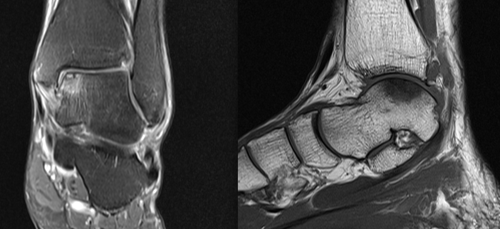
Ankle Osteochondral Defects / OCD

Osteochondral defects are a common source of pain in the ankle.  There is limited space inside the ankle joint, when you have an OCD lesion it can often feel like you have a pebble inside your shoe, with a very disconcerting crunching noise.  Once this lesion is removed and micro-fractured the results are often dramatic.  However, there is a limitation to how much pain relief can be achieved - we cannot make you better than the uninjured side.  Once damage has been done to the chondral surface - the body heals the surface with fibrocartilage, not the rock hard hyaline cartilage that you were born with.  With microfracture alone this technique is 90% effective.

I have attached imaging of some recent osteochondral lesions that have been treated at our hospital.

Case 1:


Case 2:


Case 3:


Case 4:


Case 5:


Case 6:


Case 7:


Case 8:


Case 9:


Case 10:


Case 11:


Case 12:


Case 13:


Case 14:


Case 15:


Case 16:


Case 17:


Case 18:


Case 19:


Case 20:


Case 21:


Case 22:


Case 23:


Case 24:


Case 25:


Case 26:


Case 27:


Case 28:


Case 29:


Case 30:


Case 31:


Case 32:


Case 33:


Case 34:


Case 35:


Case 36:


Case 37:


Case 38:


Case 39:


Case 40:


Case 41:


Case 42:


Case 43:


Case 44:


Case 45:


Case 46:


Case 47:


Case 48:


Case 49:


Case 50:


Case 51:


Case 52:


Case 53:


Case 54:


Case 55:


Case 56:


Case 57:


Case 58:


Case 59:


Case 60:


Case 61:


Case 62:


Case 63:


Case 64:


Case 65:


Case 66:


Case 67:


Case 68:


Case 69:


Case 70:


Case 71:


Case 72:


Case 73:


Case 74:


Case 75:


Case 76:


Case 77:


Case 78:


Case 79:


Case 80:


Case 81:


Case 82:


Case 83:


Case 84:


Case 85:


Case 86:


Case 87:


Case 88:


Case 89:


Case 90:


Case 91:


Focused Shockwave Therapy

Focused Shockwave Therapy (FSWT) is a medical treatment that uses high-energy acoustic waves to promote healing, reduce pain, and improve f...